Source: National Science Foundation
Microscopic photography is one of the most breathtaking and unconventionally beautiful forms of the artistic medium. In commemoration of the oft-overlooked genre, the Olympus BioScapes Digital Imaging Competition has recently released its selection of 2013 contest winners.
The images vary greatly in content, style, and appearance but what they do have in common is their uncanny ability to present viewers with a dynamic world that typically goes unbeknownst to the human eye. Nine images wowed judges enough to be included in the winners circle, along with one video entry.
Without further ado, here are the 2013 winners of the Digital Imaging competition:
Source: Twisted Sifter
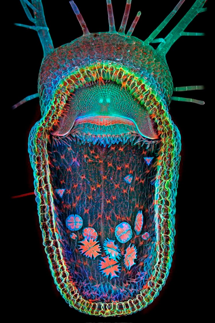
1st place: Igor Siwanowicz, HHMI Janelia Farm Research Campus, Ashburn, VA, USA. The opened trap of the humped bladderwort (Utricularia gibba), a carnivorous aquatic plant. The floating plant ingests micro invertebrates that are captured in the blink of an eye after touching one of the trigger hairs. Magnified 100x, the red in the image is the innate florescence of chlorophyll.
Source: Twisted Sifter
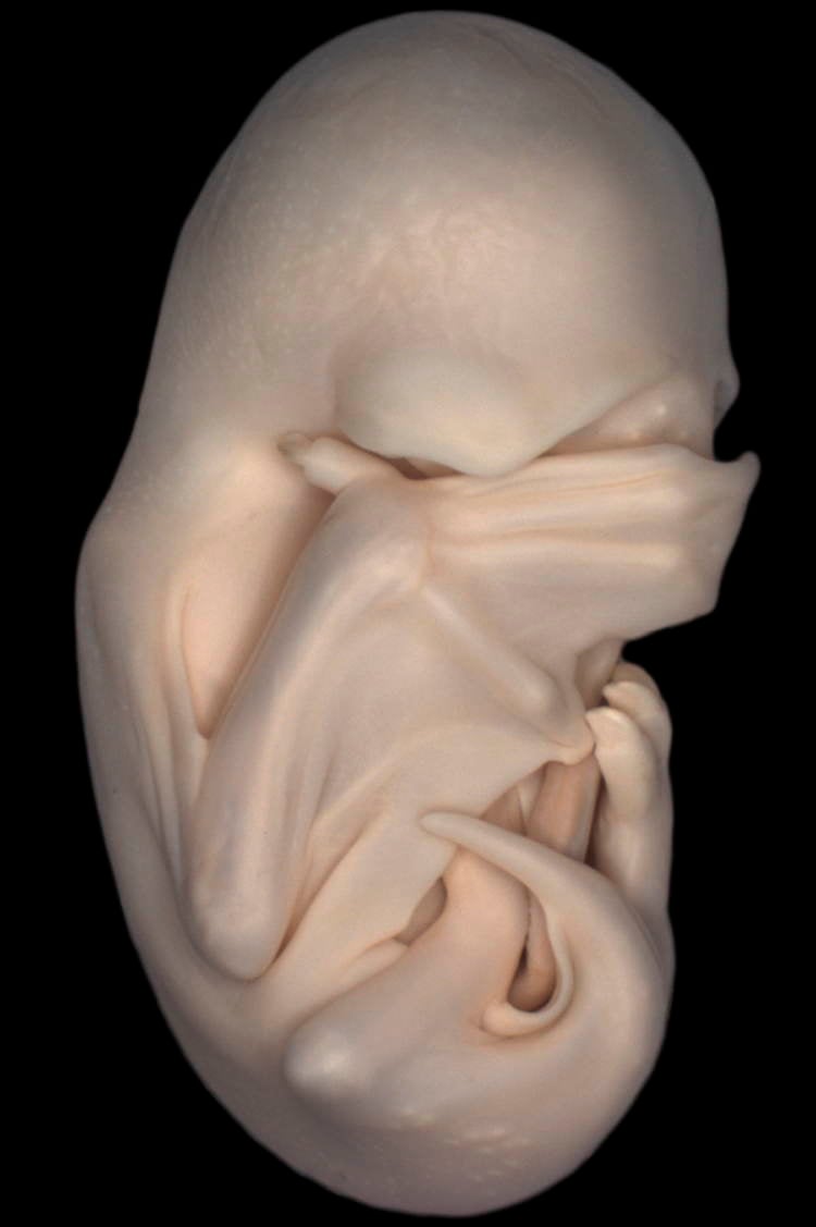
2nd place: Dorit Hockman, University of Oxford, Oxfordshire, UK. The “peek-a-boo” stage of a black mastiff bat embryo; the stage is named as such because the wings have grown to the point of covering the eyes of the bat. This image was taken under magnification of a stereo microscope.
Source: Twisted Sifter
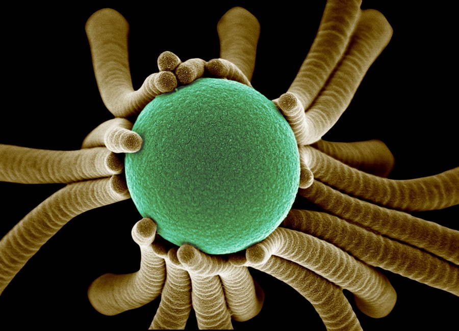
3rd place: Igor Siwanowicz, HHMI Janelia Farm Research Campus, Ashburn, VA, USA; Confocal imaging at 400x magnification. Image shows a collection of single-cell fresh water algae, desmids. The desmids can vary greatly in size, ranging from 10 microns to 1/3 of a millimeter.
Source: Twisted Sifter
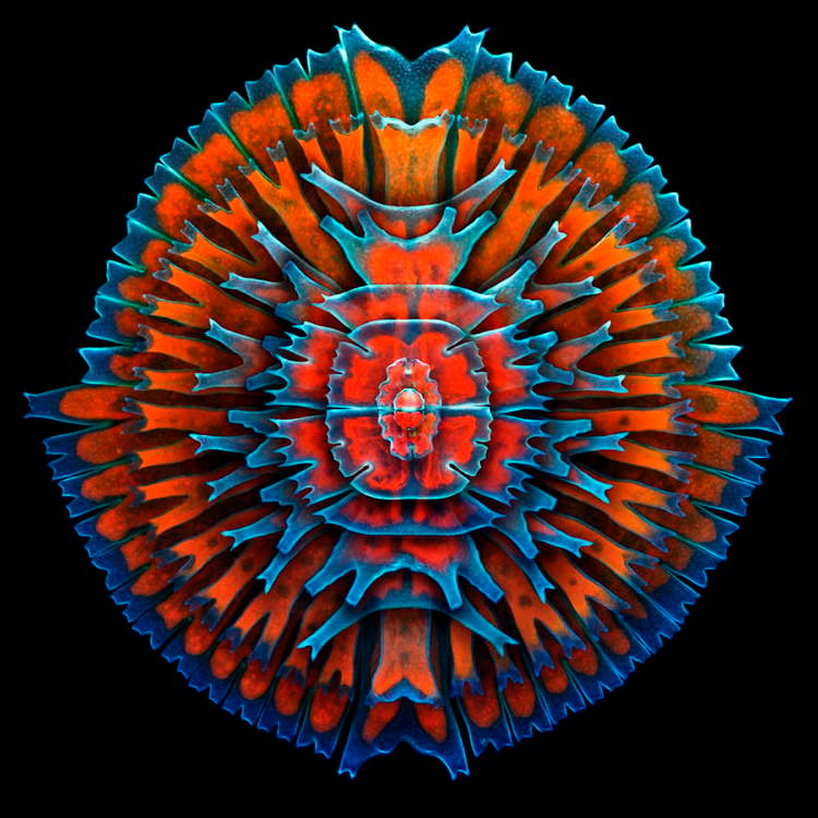
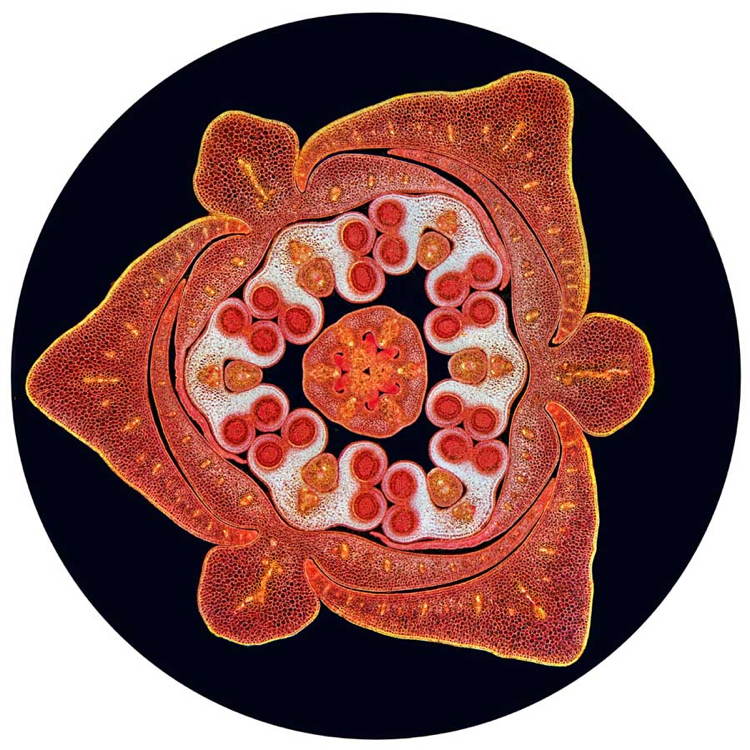
4th place: Spike Walker, Staffordshire, UK; photo of the stained transverse section of a lily flower bud, using dark field illumination.
Source: Twisted Sifter
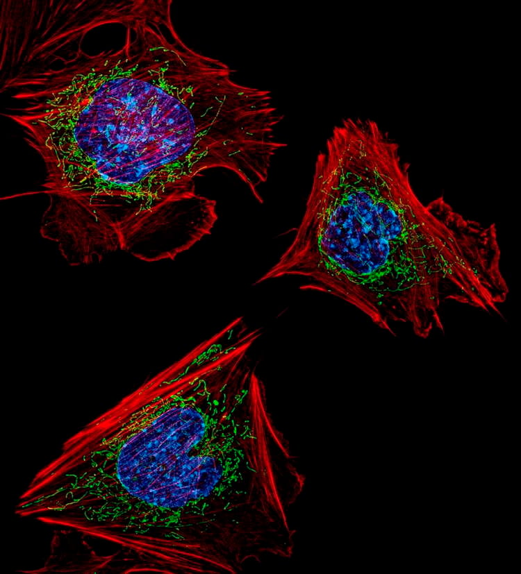
5th place: Dylan Burnette, National Institutes of Health, Bethesda, MD, USA. Structured illumination microscopy (SIM) fluorescence; image acquired with a 60x objective. Shows mouse embryonic fibroblasts showing the actin filaments (red) and DNA (blue). Also shown are the insides of mitochondria, via a green fluorescent protein (GFP) fused to a mitochondrial localization sequence.
Source: Twisted Sifter
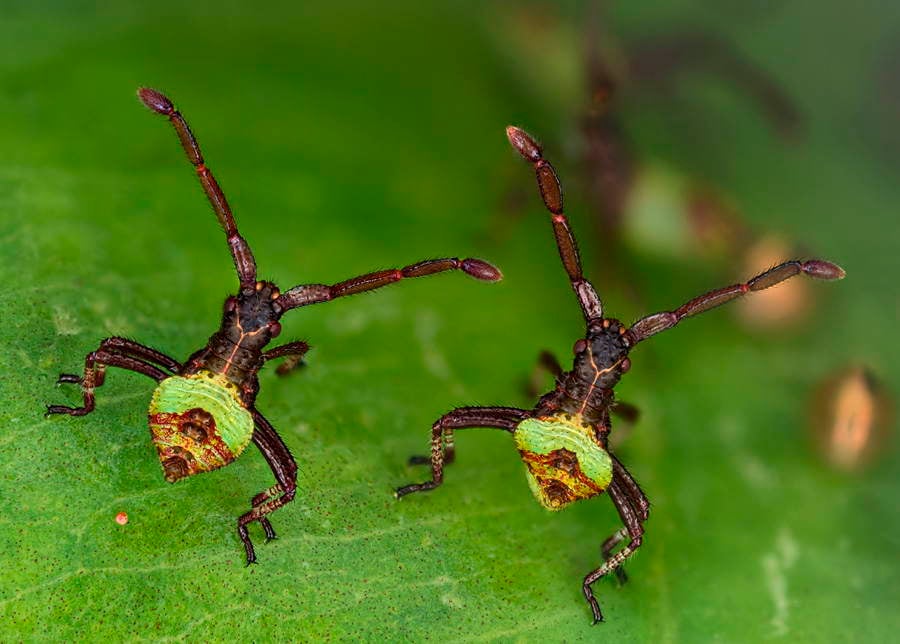
6th place: Kurt Wirz, Basel, Switzerland. Two hour old Gonocerus acuteangulatus (aka ‘brother bugs’) which are 3mm in size.
Source: Twisted Sifter
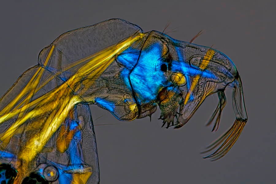
7th place: Charles Krebs, Issaquah, WA, USA. Phantom midge larva (Chaoborus) or ‘Glassworm’ The musculature which is usually clear is made visible by using polarized light for special illumination; shown at 100x magnification.
Source: Twisted Sifter
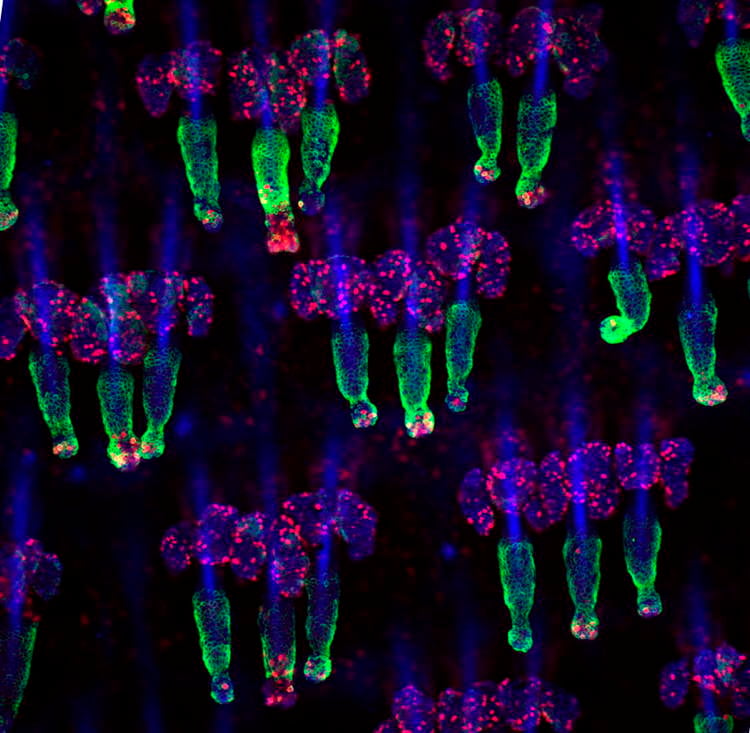
8th place: Yaron Fuchs, Howard Hughes Medical Institute/the Rockefeller University, New York, NY USA; Confocal Z-stack image. Shot with the help of technician Samara Brown. Mouse tail whole mounts stained for the K15 (green) hair follicle stem cell marker as well as Ki67 (red), which marks proliferating cells. Nuclei are marked with DAPI (blue).
Source: Twisted Sifter
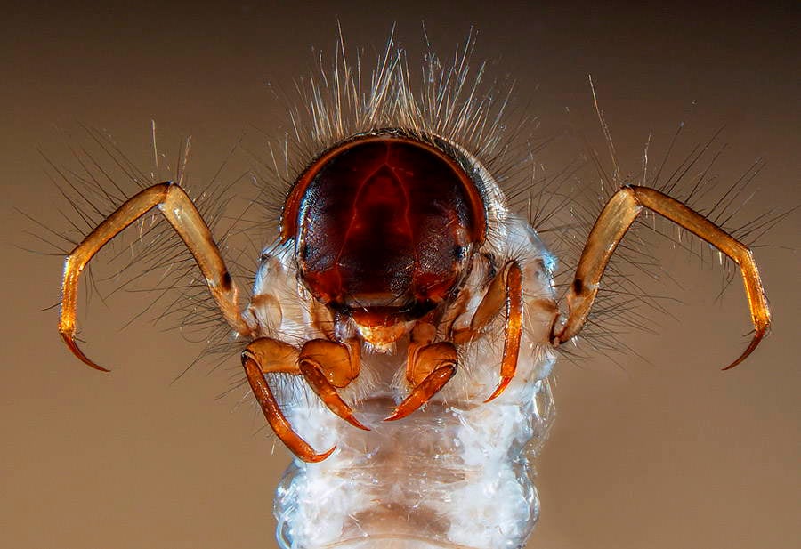
9th place: Fabrice Parais, DREAL (Regional Directorate of Environment, Planning and Housing) of Basse-Normandie, Caen, France. Upper body section and head of a caddisfly larva: Sericostoma sp; a European and North American genus of insects whose larvae live in fresh water, in gravel, stones or sand. This larva is often used to test for water pollution as its immune system is very sensitive and will cause it to die if exposed to dirty water. Stereo microscopy, 15x magnification.
For more incredible science photography, check out our gallery of the best science photos of 2013!