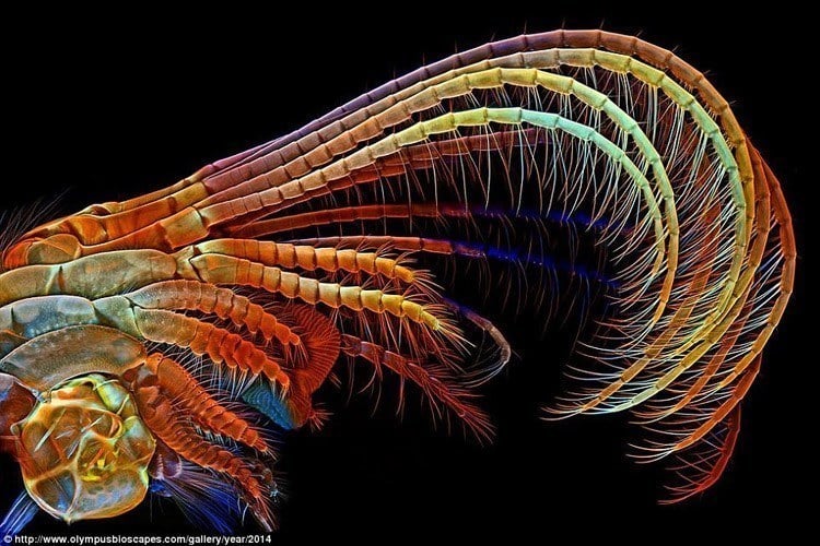
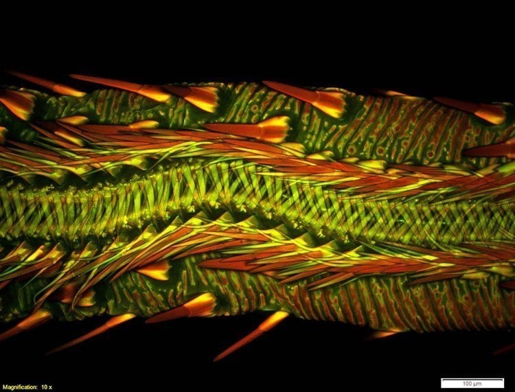
3rd Place: Dr. Igor Siwanowicz – HHMI Janelia Research Campus // Ashburn, VA, USA.
The appendages of a barnacle are used to move food for consumption. Technique: Confocal microscopy, 100x
Now in its second decade, the Olympus Bioscapes photography competition celebrates the stunning beauty of and discoveries in the field of science. But it comes with a catch: this beauty must be found beneath the lens of a microscope. Amateur and professional scientists from over 70 different countries submit thousands of entries per year in the hopes of being recognized in the competition, which is widely regarded as the world’s best showcase for this unique brand of photographic landscapes. The images that follow contain both winners and honorable mentions for 2014.
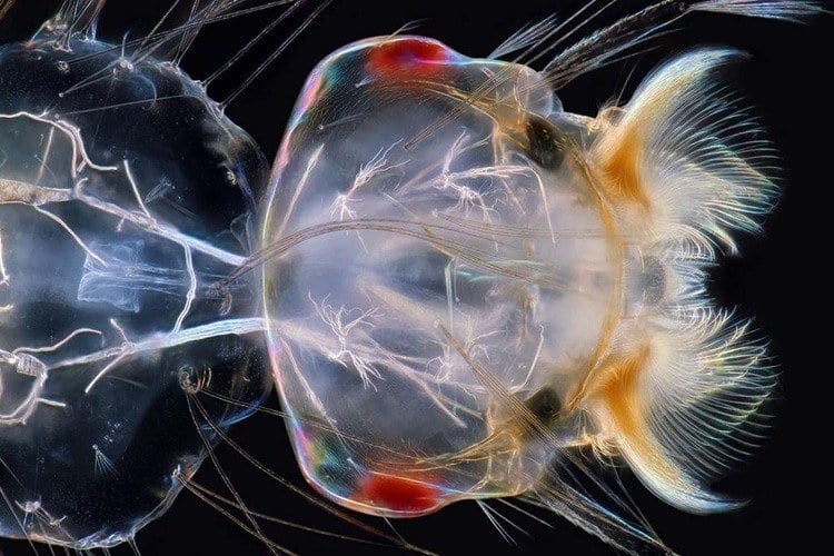
Honorable mention: Mr. Charles Krebs – Issaquah, WA, USA.
Specimen: Mosquito larva.
Technique: polarized dark field illumination, 100x
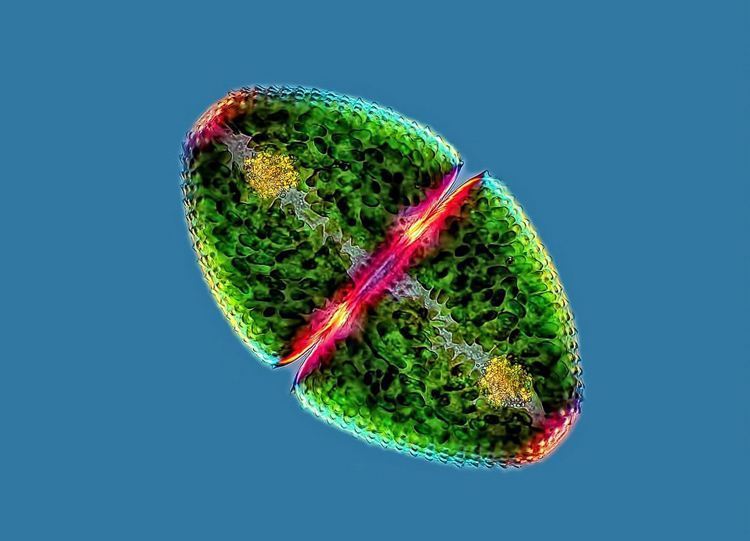
Honorable mention: Mr. Rogelio Moreno Gill – Panama City, Panama.
Micro algae from a river – with chloroplasts, isthmus and accumulation of crystals
Technique: Polarized light with image stacking
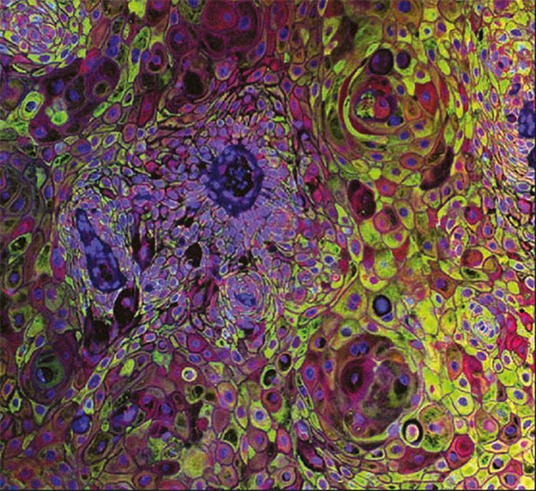
Honorable mention: Dr. Gopinath Meenakshisundaram – Institute of Medical Biology // A-Star, Singapore.
A human skin cancer cell
Technique: Confocal microscopy
Co-prizewinners: Prabha Sampath
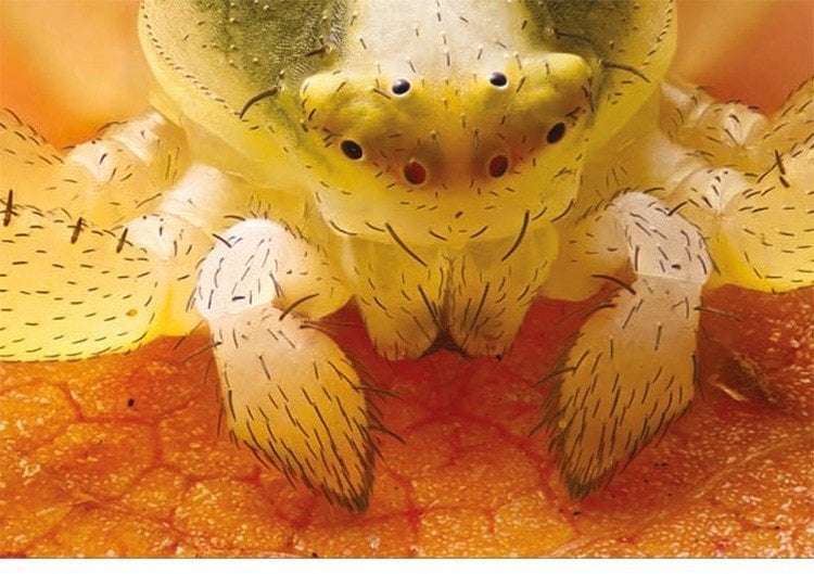
Honorable mention: Mr. Geir Drange – Asker, Norway.
Head of a young crab spider
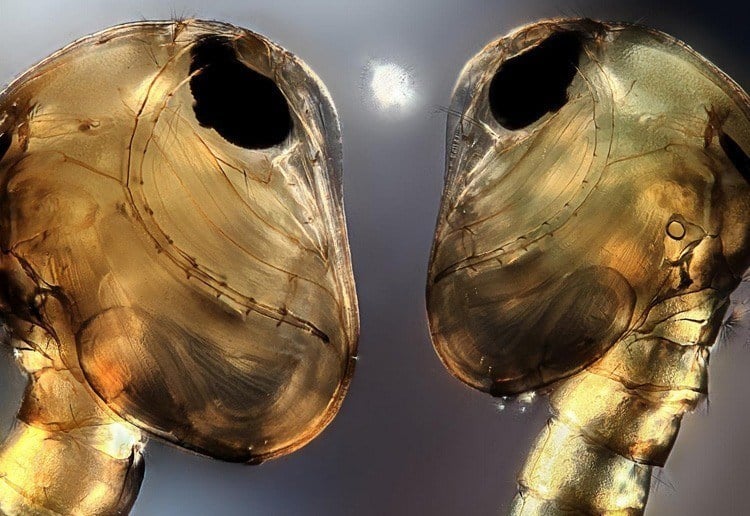
Honorable mention: Mr. Jerzy Rojkowski – Krakow, Poland.
Mosquito pupae
Technique: Differential interference contrast and image stacking, 10x
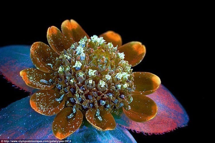
7th Place: Mr. Oleksandr Holovachov – Ekuddsvagen, Sweden.
Magnified Butter Daisy; Technique: Fluorescence
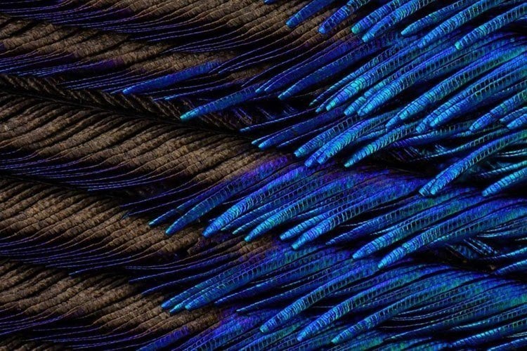
Honorable mention: Mr. Charles Krebs – Issaquah, WA, USA.
A peacock feather
Technique: Reflected light, 100x
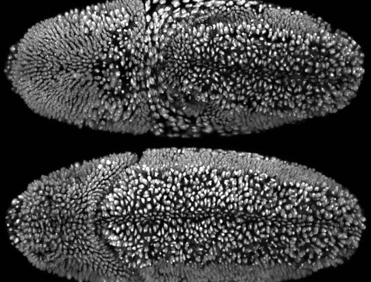
1st Place: Dr. William Lemon – HHMI Janelia Research Campus // Ashburn, VA, USA .
Embryonic development of the fruit fly. This entry is a short timelapse video showing the larvae crawling off the screen at the end. Technique: Custom-built simultaneous multi-view light sheet microscopy
Co-prizewinners: Fernando Amat and Philipp Keller
8th Place: Dr. Matthew S. Lehnert – Kent State University at Stark // North Canton, OH, USA.
The proboscis of a vampire moth
Technique: Confocal microscopy
Co-prizewinners: Ashley L. Lash