9th Place: Dr. Igor Siwanowicz – HHMI Janelia Research Campus // Ashburn, VA, USA.
Green coneheaded planthopper with a dorsal view of gears. This demonstrates that gears- which were thought to be a human invention- exist in nature.
Technique: Confocal microscopy, magnification ca. 200x
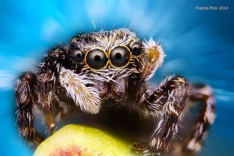
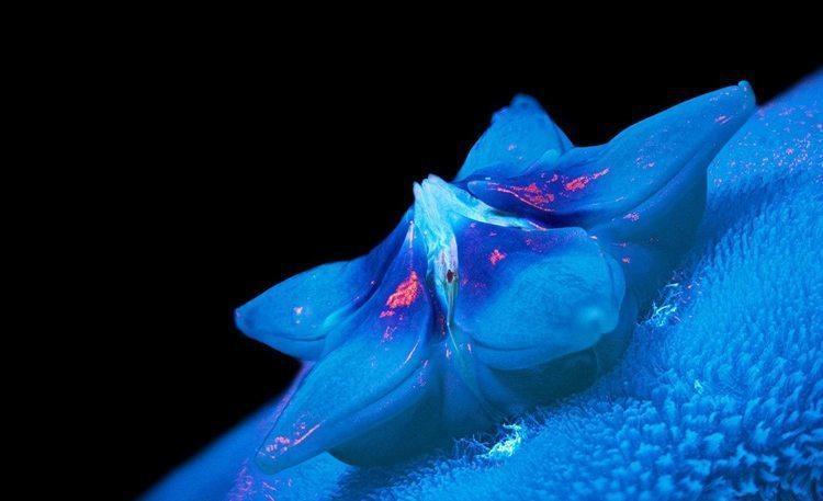
Honorable mention: Mr. Francis Prior // Liverpool, UK.
Jumping Spider
Technique: Epi-illumination
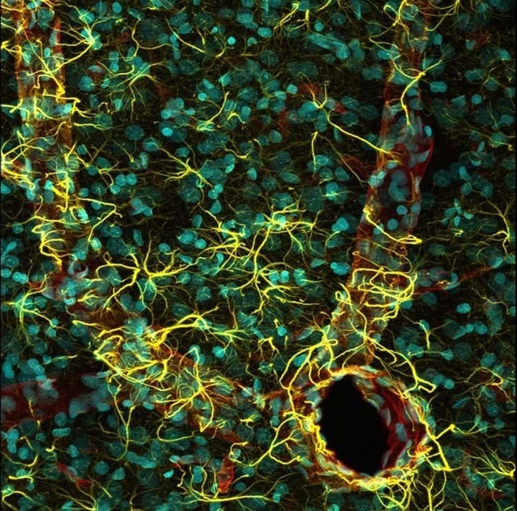
5th Place: Miss Madelyn May – Hanover, NH, USA.
The cerebral cortex of a rat
Technique: Confocal microscopy
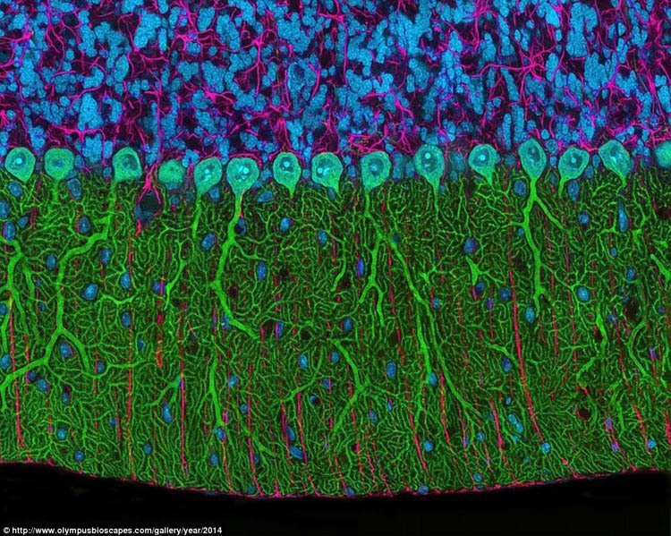
2nd Place: Mr. Thomas Deerinck – National Center for Microscopy and Imaging Research, University of California // San Diego, CA, USA.
The cerebellum of a rat
Technique: Multiphoton photography, 300x
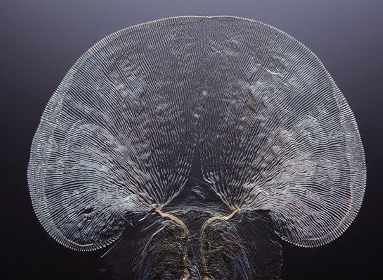
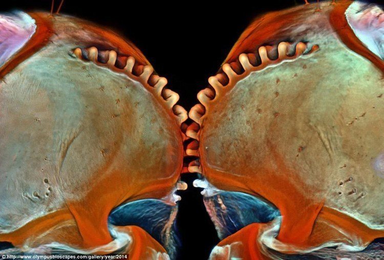
Honorable mention: Dr. David Maitland – Norfolk, UK.
The fan-like pattern of a cricket tongue
Technique: Differential interference contrast
Honorable mention: Mrs. Magdalena Turzańska – University of Wrocław // Poland.
Liverwort
Technique: Fluorescence, 125x
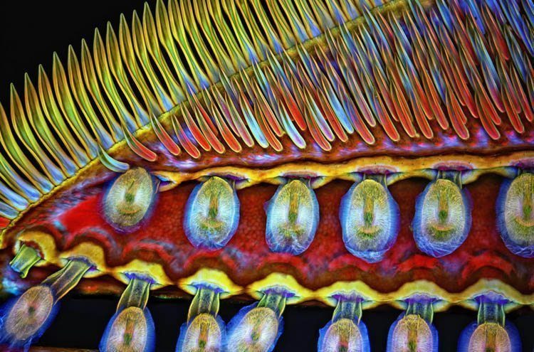
Honorable mention: Dr. Igor Siwanowicz – HHMI Janelia Research Campus // Ashburn, VA, USA.
Part of the front foot of a giant diving beetle, featuring suction cups and hollow hairs
Technique: Confocal microscopy, ca. 100x
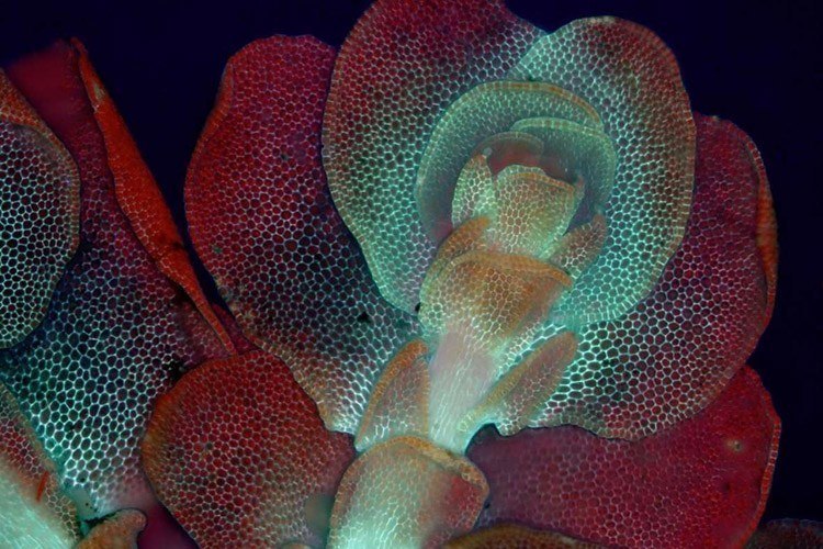
Honorable mention: Mr. Oleksandr Holovachov – Nacka, Sweden.
Wax plant flower
Technique: ultraviolet-induced visible fluorescence, image stack at 2x
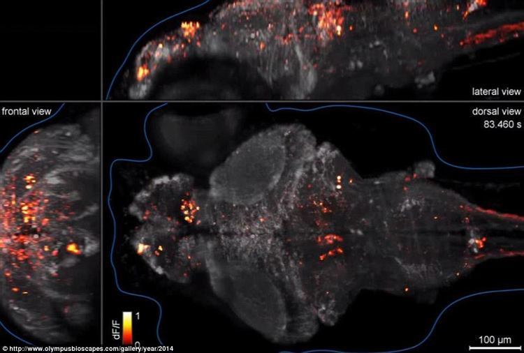
10th Place: Dr. Philipp Keller – HHMI Janelia Research Campus // Ashburn, VA, USA.
Neural activity is shown in the brain of a zebrafish. Technique: Custom-built simultaneous multi-view light sheet microscopy
Co-prizewinners: Fernando Amat and Misha Ahrens
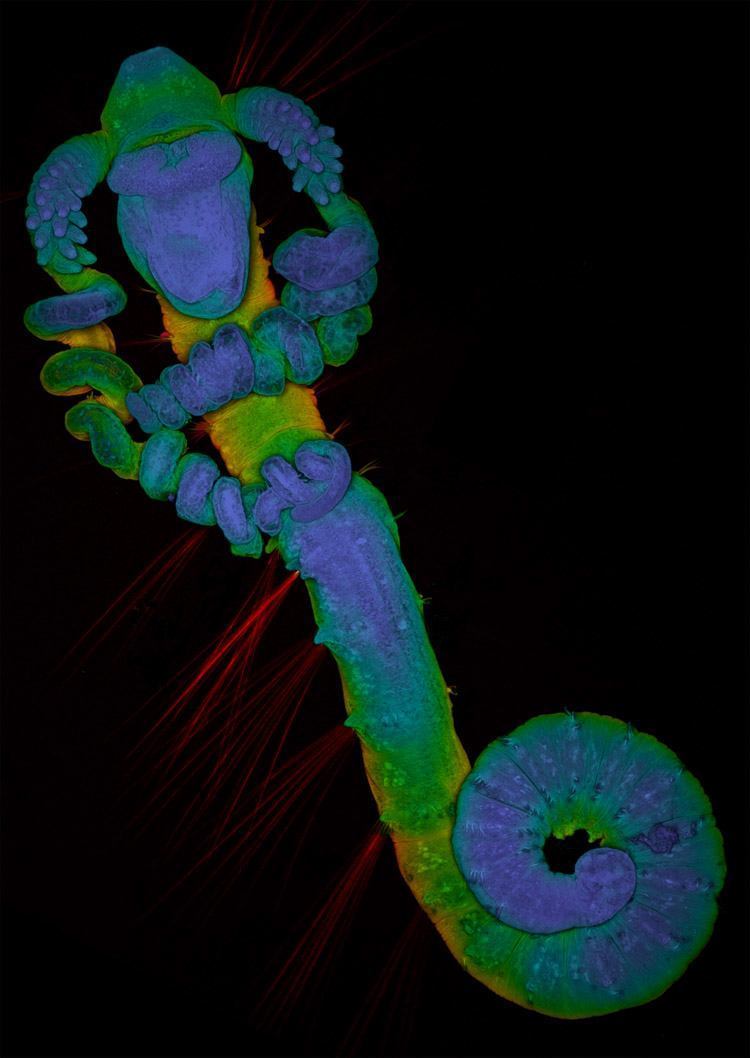
6th Place: Dr. David Johnston – Southampton General Hospital Biomedical Imaging Unit // Southampton, UK
Magelonid polychaete worm larva.
Technique: Confocal microscopy using a 10x objective
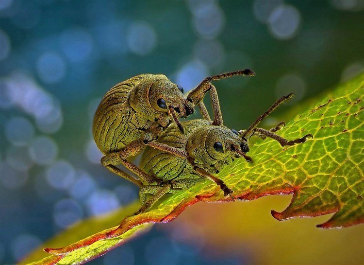
4th Place: Dr. Csaba Pintér – Keszthely, Hungary.
Phyllobius roboretanus weevils
Technique: Stereo microscopy
All images can be found on the Olympus Bioscapes competition page.