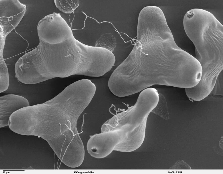
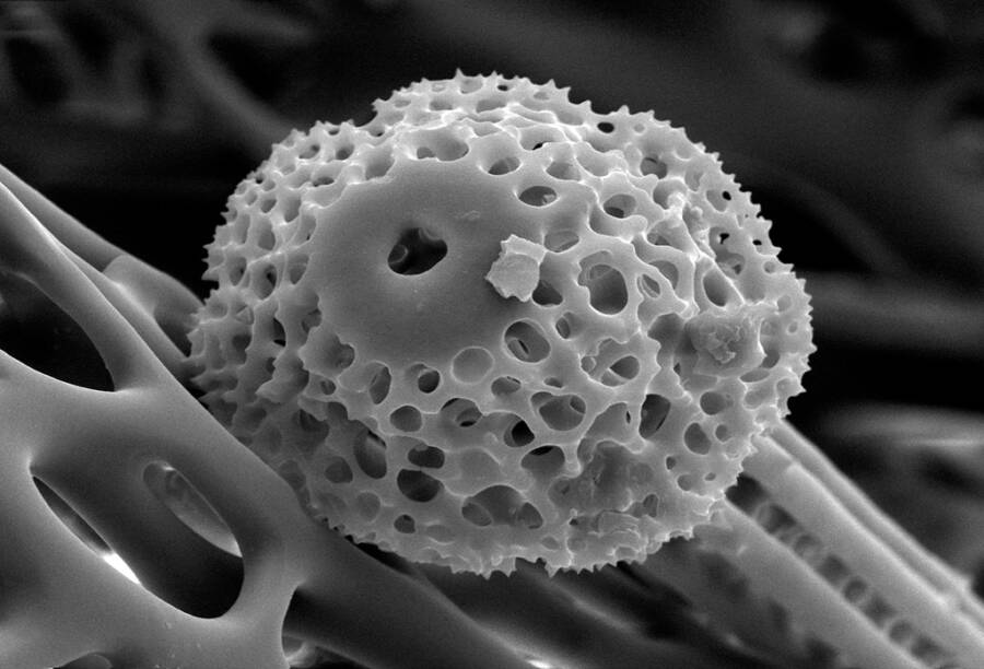
These electron microscope images show what life is like in the invisible world of viruses, bacteria, and pollen — and it’s both surreal and spooky.
The microscopic world is an endlessly fascinating place, and thanks to technological advances over the last 90 years, we can now see things at incredibly high magnification through these electron microscope photos.
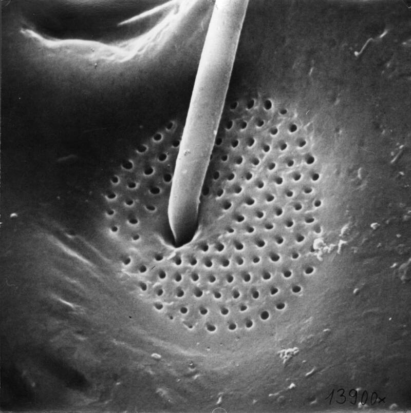
Scanning electron microscopes (SEM) show us the invisible world of microorganisms by combining a variety of signals that are then scanned through a focused beam of high-energy electrons spread across a specimen. The electrons scatter, and the microscope uses this scattering to recreate an image.
Such electron interactions give us information like topography, texture, chemical composition, and the orientation of those materials within the sample.
Combining these informational signals into one image provides a somewhat two-dimensional, black and white photo; sometimes artificially colorized. The magnification can range from 10x – 300,000x, with some microscopes even scanning up to 500,000x.
Electron microscopes are widely used in science and engineering, and the range of different techniques its users can employ is extremely versatile. In addition, SEMs have made possible nearly all recent advances in the material science industries — from aerospace and chemistry to electronics and energy usage.


And if you liked this post, be sure to check out these popular posts:


Inside The History Of The Scanning Electron Microscope
Since its invention in 1931 and the launch of its commercial availability in 1965, SEM technology has become a staple of academic research.
Max Knoll and Ernst Ruska of the Berlin Technische Hochschule were the first ones to overcome the problem of resolution limits in earlier microscopes. Their earliest prototypes proved that electron beams could be tamed to provide clearer images in a microscope.
Progress in electron lens technology also minimized defects that made for a clearer picture. In turn, this led to greater resolutions. Once the electron technology was in place, electron microscopy would advance through the use of brighter electron guns and improved vacuum systems.

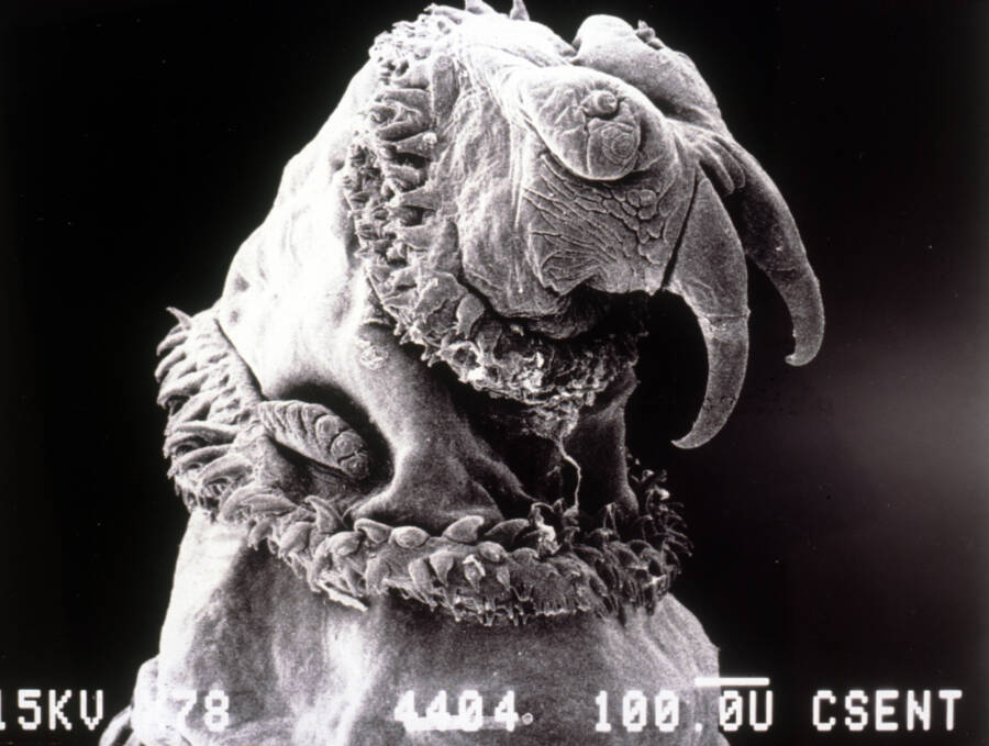
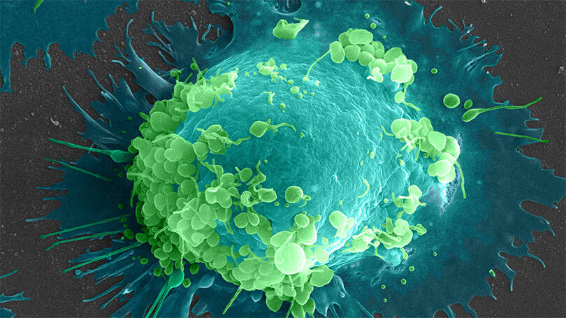
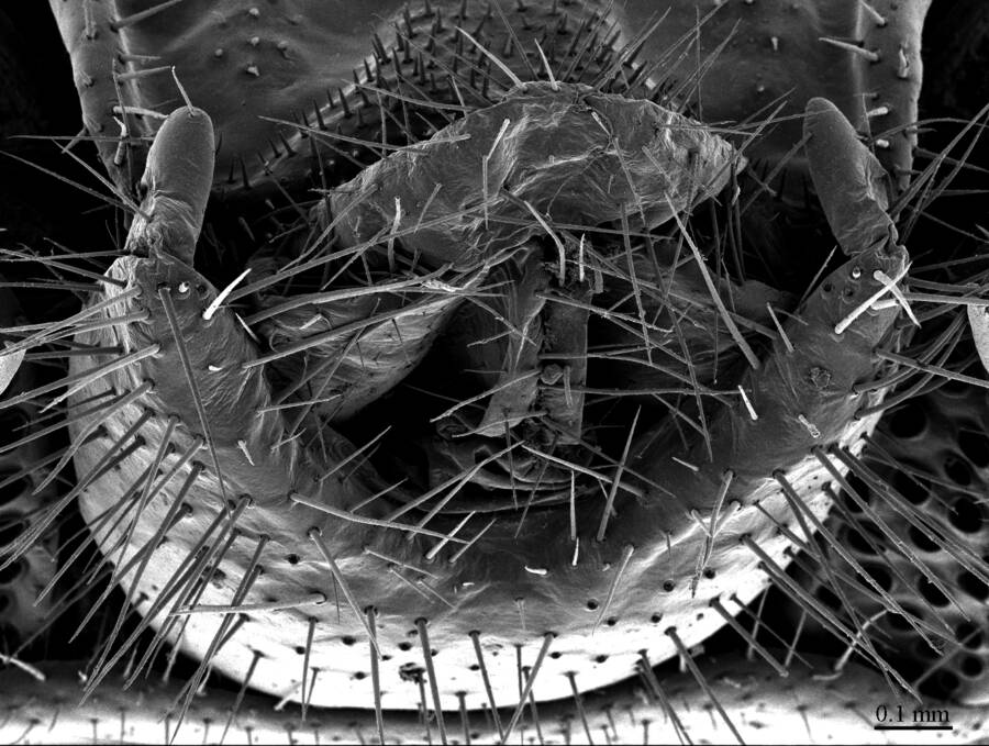
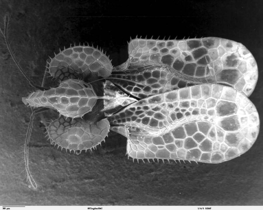
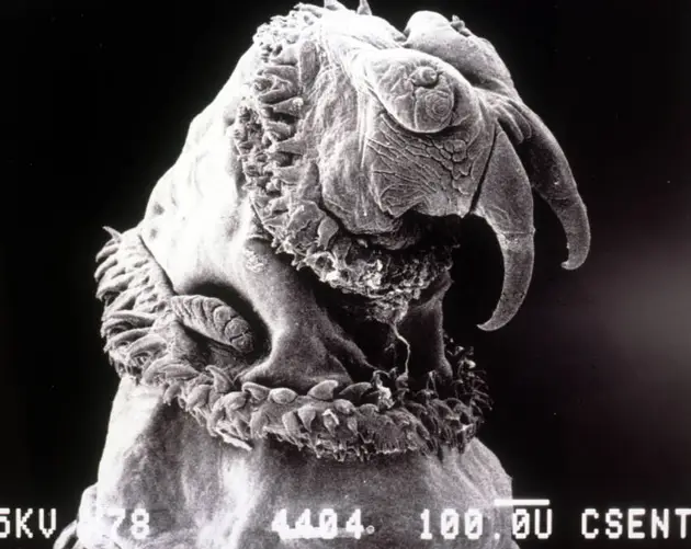
ImgurImages taken with a scanning electron microscope are helping scientists to better understand viruses and diseases. Pictured here, though, is a mite.
Once commercialized, scientists behind the scenes at many household-name companies began tweaking the tech and selling SEMs commercially. RCA was the first in North America to pedal electromagnetic lenses. General Electric tried to compete with them, selling their electrostatic electron microscopes. Philips, Hitachi, and Toshiba also played roles in the development process.
All sorts of electron microscopes hit the market; some for the beginner and others for people with advanced knowledge. The engineers, of course, wanted to achieve the highest resolution possible. But they had to take into account what would sell most readily to the everyday consumer looking for ease of use.
The ultimate goal of this technology was to eventually get to atomic-level resolutions. However, this wouldn't come to pass until the 1980s.
Why These Innovative Tools Are Useful
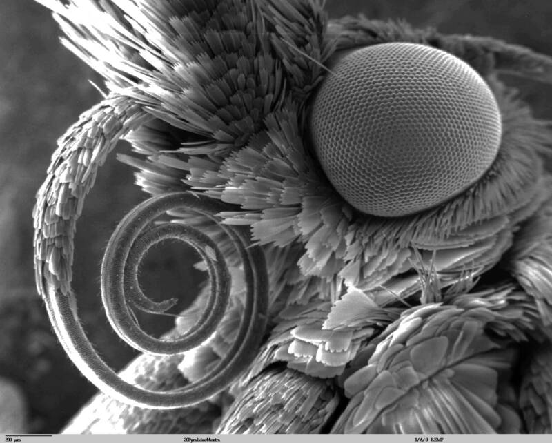
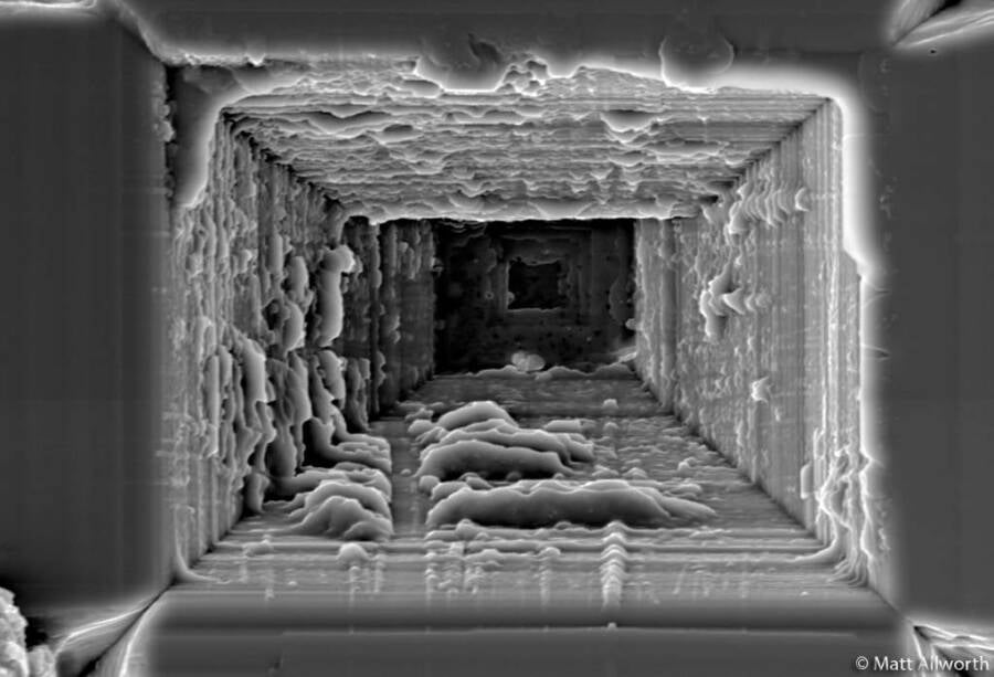
In addition to just looking really cool, SEMs are utilized in several industries: medical, industrial, and research — just to name a few. Microchip production relies heavily on scanning electron microscopes, as does semiconductor inspection.
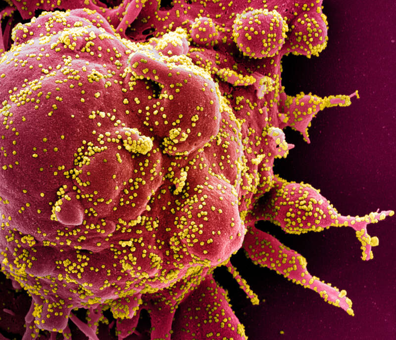
Medical laboratories use electron microscopes to examine both biological and non-biological specimens and to identify new viruses and diseases. Furthermore, SEMs are able to test new vaccines and treatments.
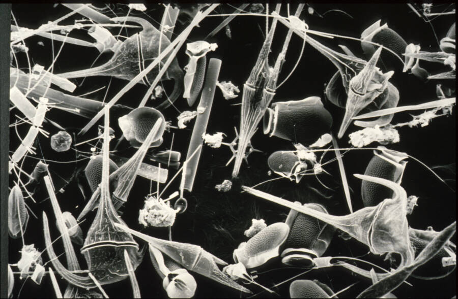
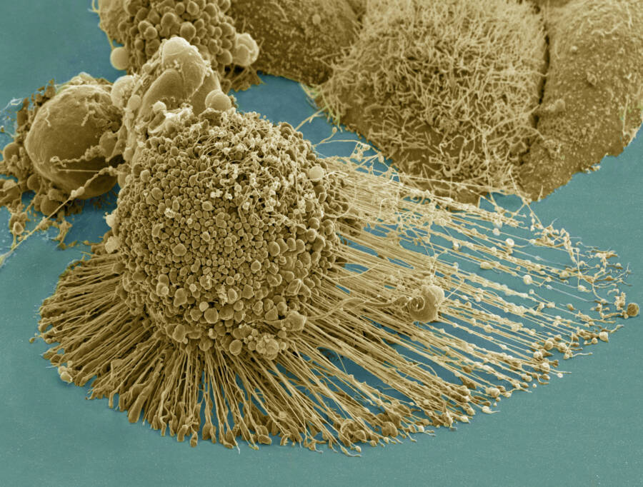
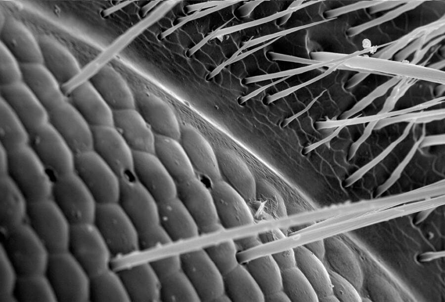
Wikimedia CommonsA sample of microorganisms taken from the Derwent River water in south Australia.
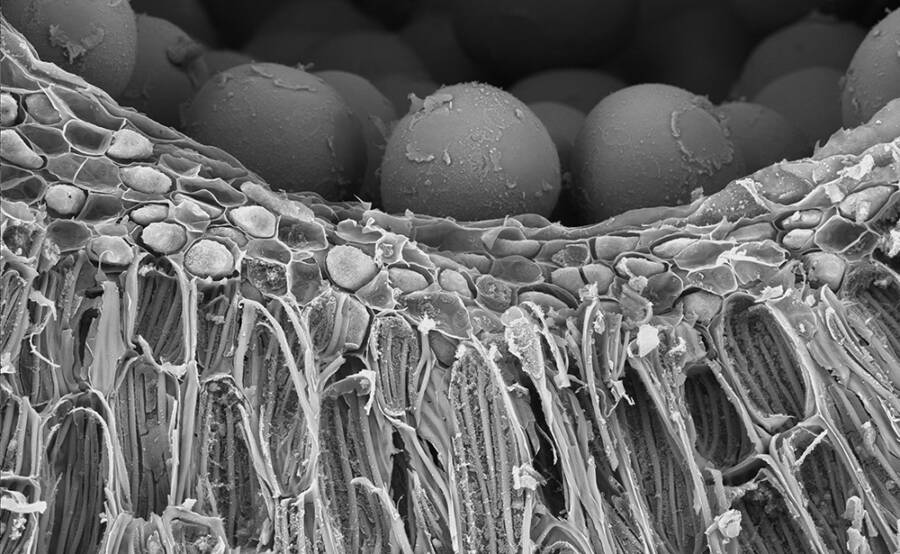
Researchers can even investigate the detailed structures of tissues and cells at this scale, counting individual particles within samples.
SEMs also help identify early human artifacts and assist in dating historic ruins.
This technology is also useful in soil quality control in the areas of farming and agriculture. Electron microscopes identify compositional differences and weathering processes on all kinds of rocks and minerals.
Even the justice system uses data from electron microscope photos in courtrooms. Forensic scientists use SEMs to detect gunshot residue, examine bullet markings, identify paint and fiber composition — even handwriting analysis.
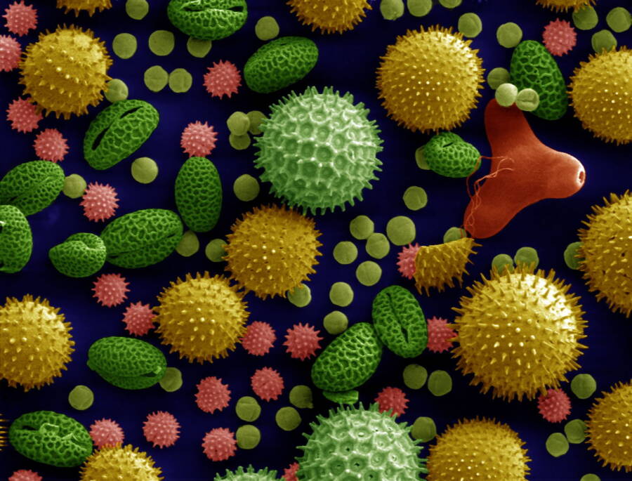
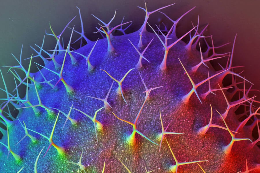
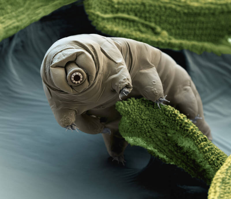
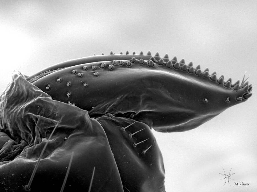
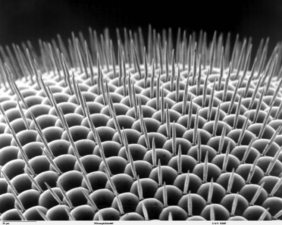
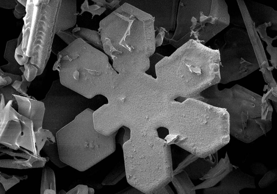
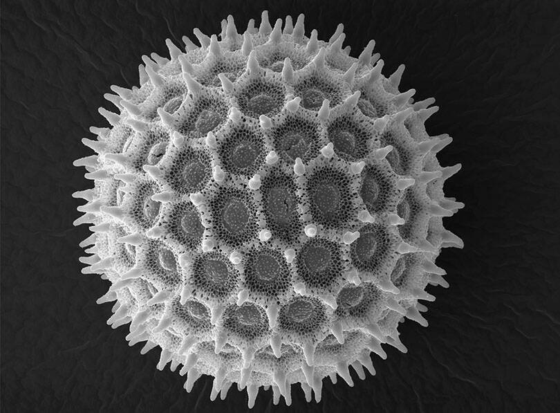
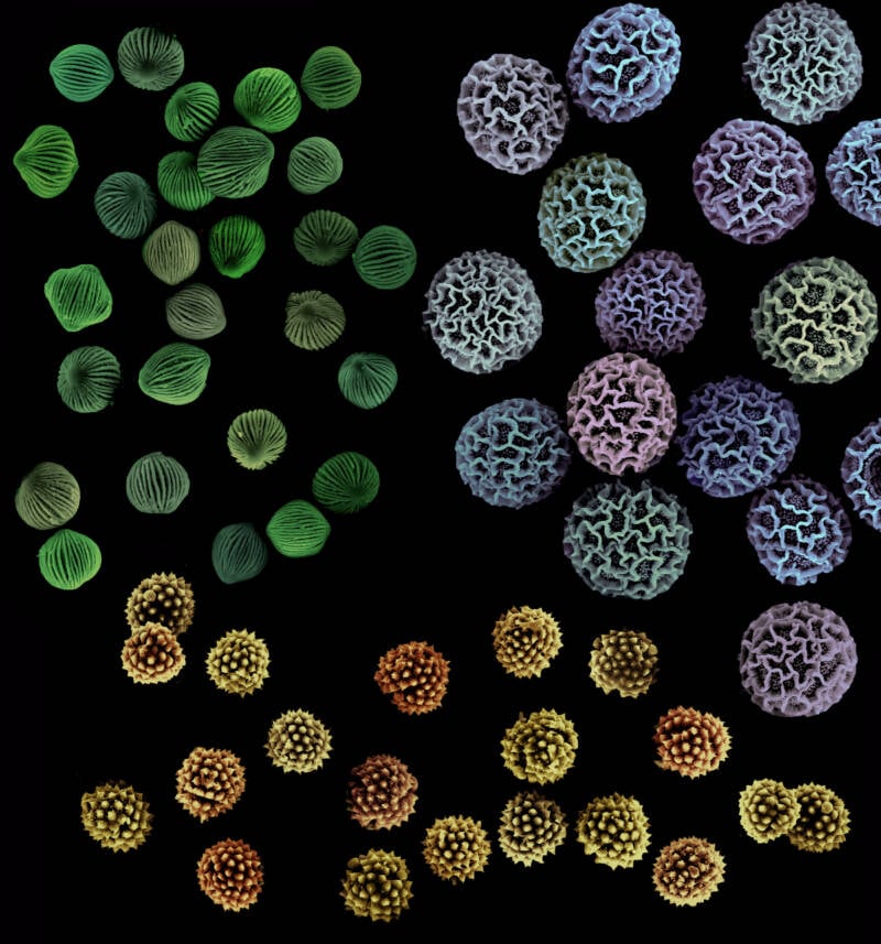
Of course, as is clear by these scanning electron microscope photos, not all uses are strictly practical in nature. Micrographs made by SEMs are sometimes used to make digital artworks. Microscopic materials can become diverse landscapes — both alien or eerily familiar.
After this look at these electron microscope images, check out these 27 jaw-dropping science photos from 2020 that you may have missed. Then, find out how researchers determined the diet of the ancient builders of Stonehenge consisted of parasite-infested meat.